You’re offline. This is a read only version of the page.
Search
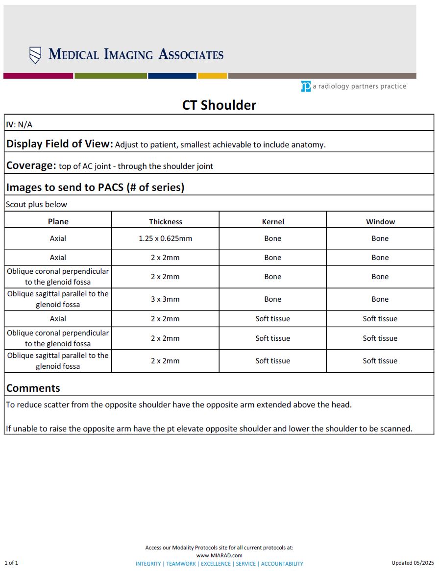
CT Shoulder
DOWNLOAD PDF