


MR Ankle
Common Histories / Key Words:
Tendonitis/Tendon Tear, Ligament Tear/Sprain, Ankle Pain, Fracture, Trauma/Injury, Osteochondral Lesion/Defect, Plantar Fasciitis
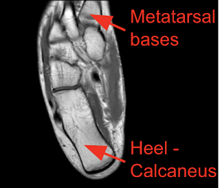
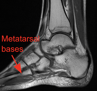
COVERAGE: An ankle MRI should extend from the soft tissues posterior and plantar to the heel through the metatarsal bases as depicted below:
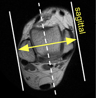
SAGITTAL - Imaging to extend from the medial soft tissues to lateral soft tissues
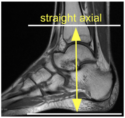
AXIAL - Imaging to include the distal tibial metaphysis through the plantar soft tissues
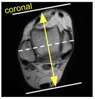
CORONAL - Imaging to include the soft tissues posterior to the heel through the metatarsal bases
FOV:16x16
Matrix: 256x224
Slice Thickness: 3.5/0.5
ROUTINE
■ Sag T1 and T2FS (or STIR)
■ Ax PD (No FS) and T2 FS
■ Cor T2 FS
ACHILLES TENDON
■ Sag T1 and T2FS (or STIR)
■ Ax PD (No FS) and T2 FS
Please see
MR MASS - INFECTION - ALL JOINT/EXTREMITIES
link for post IV contrast protocol
METAL ARTIFACT - STIR sequences may be substituted in place of T2 FS when metal artifact is present in attempt to improve signal and image quality. Please see link below for additional suggestions.
>MR METAL ARTIFACT