


MR Elbow
ELBOW
Common Histories / Key Words: Ulnar Nerve Impingement, Epicondylitis, Biceps Tear, R/O Fracture, Collateral Ligament Tear, Loose Bodies.
POSITIONING: Supine with upper extremity next to patient's side. Place arm into supination with palm up. Sandbags may be placed on the palm to help reduce motion and retain position.
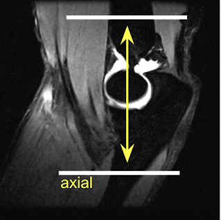
AXIAL - 4 cm above the joint and through the bicipital tuberosity of the radius (bicep's tendon insertion)
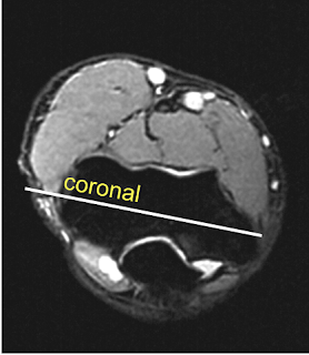
CORONAL - Use axial image to make slices parallel to the horizontal line drawn between the humeral epicondyles.
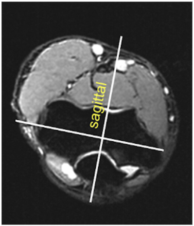
SAGITTAL - Use axial image to make slices perpendicular to the horizontal line drawn between humeral epicondyles.
FOV: 14x14 (axial FOV can decrease to 12x12 in most cases)
Matrix: 256x224
Slice Thickness / Spacing: 3.5/0.5 (axial images 4.0/1.0 for coverage if necessary)
ROUTINE
■ Ax T1, T2 FS
■ Cor T1, PD FS
■ Sag T2 FS
ARTHROGRAM
■ Ax T1 (NO FS), T2 FS
■ Cor T1 FS, T2 FS
■ Sag T1 FS, T2 FS
Please see
MR MASS - INFECTION - ALL JOINT/EXTREMITIES
link for post IV contrast protocol
METAL ARTIFACT - STIR sequences may be substituted in place of T2 FS when metal artifact is present in attempt to improve signal and image quality. Please see link below for additional suggestions.
>
MR METAL ARTIFACT