


MR Hand - Finger - Thumb
MR HAND - FINGER - THUMB
ROUTINE HAND - FINGER - THUMB
■ Ax T1, T2 FS
■ Cor T1, PD FS
■ Sag T2 FS
MASS - INFECTION - ARTHRITIS
Place marker over area of interest when applicable.
■ PRE-CONTRAST
■ Axial T1, T2 FS (If there is a MASS - add an Axial T1 FS)
■ Coronal T1, T2 FS
■ Sagittal T1, T2 FS
■ POST-CONTRAST
■ Axial T1 FS, Cor T1 FS, Sag T1 FS
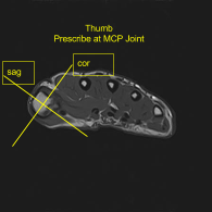
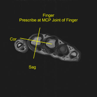
When imaging a finger or thumb specifically:
■ Three planes are performed relative to the joint in question, NOT the MRI table.
■ FOV should be selected appropriate for the body part, often 8 to 10 cm for finger, but may be larger for the whole hand.